In the ever-evolving field of medical imaging, advancements in technology are constantly redefining the possibilities of diagnostics. A notable breakthrough that has transformed the way we analyze internal structures is 3D X-ray technology.
In this blog post, we will delve into the intricacies of 3D X-ray imaging, exploring its principles, applications, and the revolutionary impact it has had on various fields.
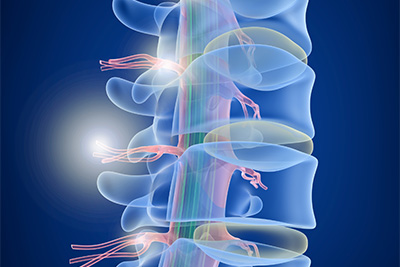
The technology of 3D X-ray, or cone beam computed tomography (CBCT), differs from traditional X-ray methods in that it captures several images from various angles around the object being scanned. This multidirectional approach allows for the creation of a three-dimensional reconstruction, providing a comprehensive and detailed view of the scanned area.
CBCT has become a cornerstone in dental imaging, offering dentists and oral surgeons a comprehensive view of the oral and maxillofacial regions. From precise implant planning to root canal assessments, 3D X-ray technology enhances diagnostic accuracy and facilitates better treatment outcomes.
When it comes to evaluating musculoskeletal structures in orthopedics, CBCT is essential for providing unmatched detail. Using 3D imaging to plan preoperatively, accurately detect fractures, and evaluate joint problems, surgeons can create a roadmap for complex orthopedic treatments.
The versatility of 3D X-ray technology extends to interventional radiology, guiding minimally invasive procedures with enhanced precision. Real-time 3D imaging aids in catheter placements, tumor ablations, and vascular interventions, elevating the standards of patient care.
Three-dimensional X-ray technology offers a better sense of depth than standard X-rays, which give a flat, single-plane image. This is especially useful for studying regions with overlapping features or intricate anatomical processes.
Overlapping structures can be a challenge in traditional X-rays, leading to potential diagnostic limitations. 3D imaging minimizes this issue, offering clearer visualizations of intricate anatomies.
When planning an operation or providing intraoperative assistance, 3D X-ray equipment is helpful to surgeons. More accurate surgical procedures are made possible by detailed 3D reconstructions, which help visualize important structures.
Although there are many advantages to 3D X-ray technology, worries regarding radiation exposure still exist. Ongoing technological developments, however, aim to optimize radiation dosages so that the advantages of diagnosis exceed any possible hazards.
Future developments in 3D X-ray imaging could lead to even more advanced diagnostics as technology advances. Applications like artificial intelligence algorithms for automated picture analysis and virtual reality integration for surgical planning are being investigated by researchers.
The use of 3D X-ray technology has revolutionized medical diagnostics, providing unprecedented precision and depth. This advancement has had a transformative impact across various medical specialties, equipping healthcare professionals with the necessary tools to make informed decisions and deliver optimal patient care. As we continue to navigate the rapidly evolving landscape of medical imaging, 3D X-ray technology serves as a testament to the power of innovation in advancing healthcare outcomes.
At Suburban Medical Group, we offer on-site X-ray services to provide fast, accurate imaging for a more precise diagnosis. Whether evaluating fractures, infections, or other conditions, our state-of-the-art X-ray technology ensures you receive timely and effective care all without needing an additional trip to another facility.